If a small hole remains where an implant was removed, is it safe to simply leave it alone?
If water seems to pass toward your nose when you drink, you may want to consider the possibility of an oroantral fistula.
Even when the visible opening looks small, the condition beneath the surface can be very different.
Let’s look at what needs to be checked during diagnosis and treatment, using a real oroantral fistula closure case.
Key Takeaways
An oroantral fistula is a condition in which an abnormal passage remains between the mouth and the maxillary sinus after an upper molar extraction or implant removal.
Even when the opening visible in the mouth is only 1-2mm, a CT scan can sometimes reveal bone loss of 15mm or more, which means a visual check alone isn’t enough to assess the condition.
That’s why, when considering oroantral fistula closure surgery, it’s important to evaluate not just the size of the fistula but also the surrounding bone and the condition of the maxillary sinus.
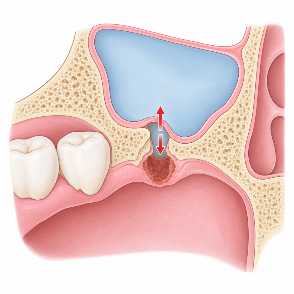
What Is an Oroantral Fistula?
An oroantral fistula refers to a persistent abnormal opening between the mouth and the maxillary sinus.
The maxillary sinus is an air-filled space located beside the nose, inside the upper jawbone.
Because the roots of the upper molars can sit close to the floor of the maxillary sinus, an extraction or implant procedure can sometimes connect the mouth and the sinus.
According to international literature, an oroantral fistula develops in about 0.3-3.8% of upper molar extractions.
If the opening is small early on, it can sometimes heal on its own, but if the passage persists, it may progress into an oroantral fistula.
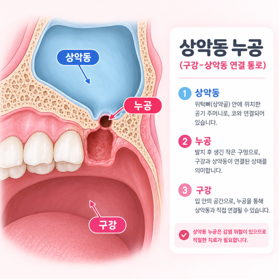
✅ A feeling of air passing between the nose and mouth
✅ A sensation of water passing toward the nose when drinking
✅ An extraction or implant removal site that won’t heal
✅ Recurring discomfort in the area

Because the roots of the upper molars can be close to the maxillary sinus, the mouth and sinus may become connected after an extraction — this is known as an oroantral communication.
The Korea Disease Control and Prevention Agency’s National Health Information Portal describes this in its entry on tooth extraction.
However, these symptoms alone cannot confirm an oroantral fistula, so it’s important to check the condition through an oral exam and any necessary imaging tests.
A Fistula Remained After Implant Removal — What Did the Real Case Reveal?
This case involves a patient who had previously received implant treatment along with a sinus lift in the upper molar area.
After a problem developed with the implant, it was removed about two years ago, but the patient came in with a fistula still present between the removal site and the maxillary sinus.
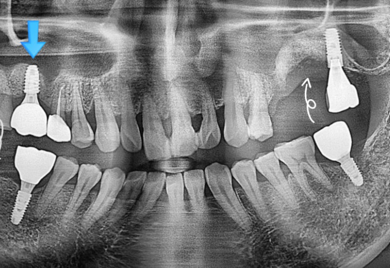
A panoramic X-ray was taken first to check the previous implant position and surrounding condition.
However, a panoramic image alone can have limits in pinpointing the exact extent of the fistula and the internal bone loss.
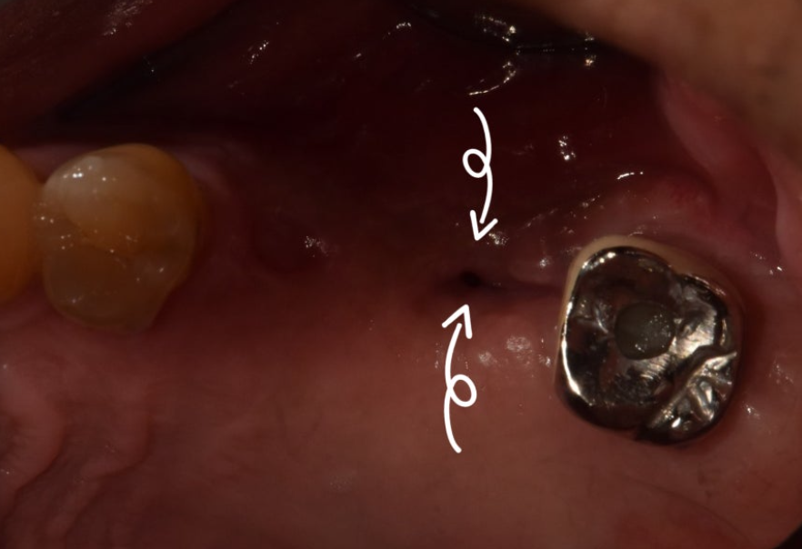
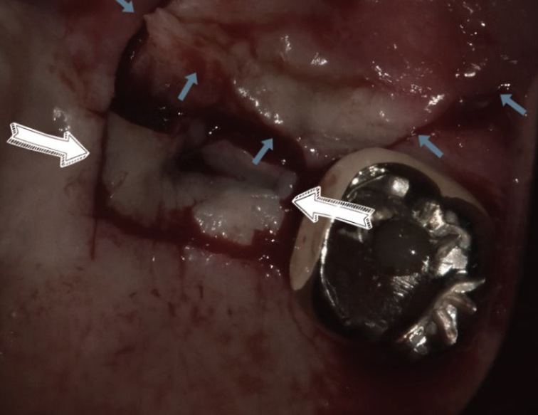
On direct oral examination, the fistula appeared to be about 1-2mm in size.
To the naked eye, it looked like a relatively small opening.
But was the internal condition really that small too?
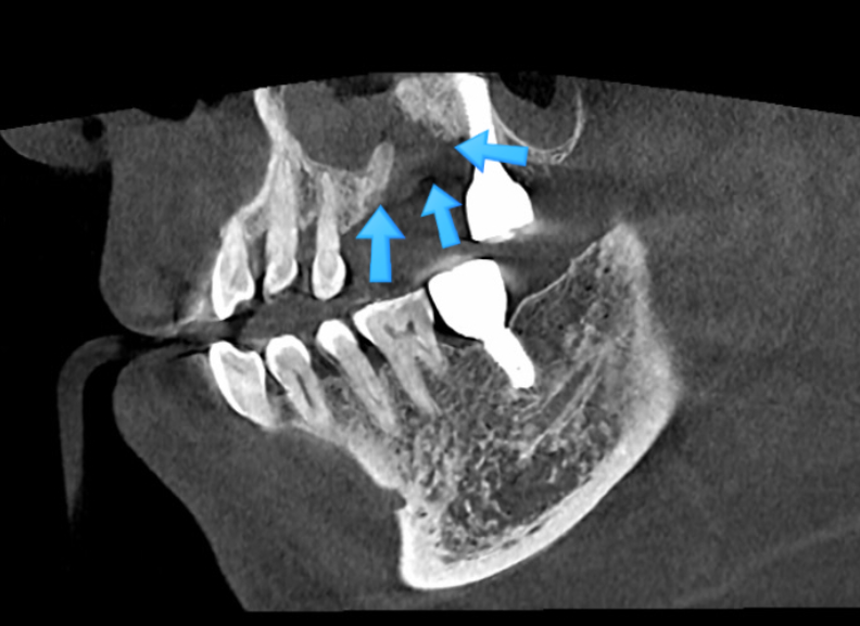
A CT scan was taken to further evaluate the internal condition, and it revealed bone loss of about 15mm beneath the gum.
There was a clear difference between the small opening visible in the mouth and the extent of the defect found internally.
The fistula visible in the mouth was only about 1-2mm, but the CT scan showed bone loss extending to about 15mm.
This case illustrates how the outward appearance and the internal condition can differ.
An international study introduced in the Journal of the Korean Dental Association reported that among 308 patients with oroantral fistulas, 187 (60.7%) underwent closure using a buccal advancement flap, and the fistula recurred in 12.8% of those cases.
Even after a closure method is chosen, recurrence is possible, which is why a thorough evaluation of the maxillary sinus and surrounding tissue is important.
How Is Oroantral Fistula Closure Surgery Performed?
Oroantral fistula closure surgery is a treatment performed to seal a persistent passage between the mouth and the maxillary sinus.
The method used can vary depending on the fistula’s location and size, the extent of bone loss, and the condition of the surrounding tissue.
- Assess the condition of the tissue around the fistula
- Check for inflammation inside the maxillary sinus
- Select a closure method suited to the defect
- Reposition and suture a tissue flap, using PRF if needed
- Monitor progress and manage recovery
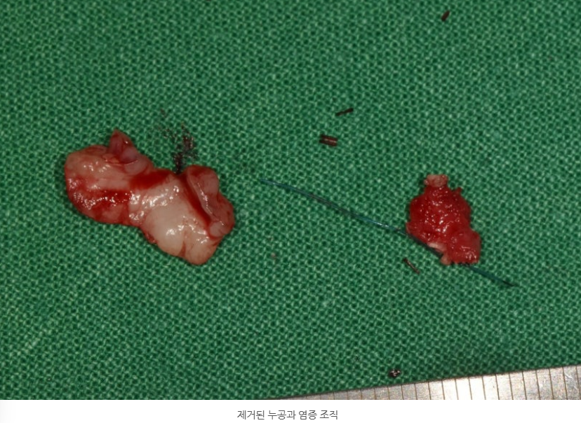
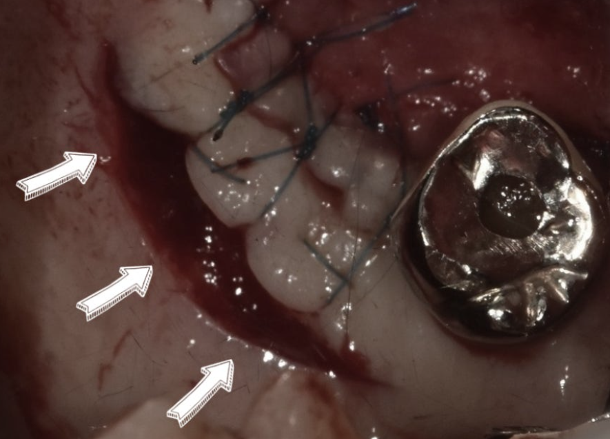
In this case, the tissue around the fistula was cleaned up, and a buccal flap on the cheek side was repositioned to cover and suture the defect.
Considering the patient’s condition, PRF and palatal tissue were also used together during suturing.
PRF is a material derived from the patient’s own blood, using platelet and fibrin components, and it may be used as needed during oral surgery procedures.
Not every oroantral fistula closure surgery uses the same method or materials.
It’s important to check the fistula’s size and location, the amount of surrounding tissue, and the condition of the maxillary sinus, then decide on a treatment approach suited to each individual case.
| Factor Checked | Impact on Treatment Plan |
|---|---|
| Fistula size and location | Determines closure extent and method |
| Degree of bone loss | Whether additional treatment is needed |
| Inflammation inside the sinus | Whether prior treatment is needed |
| Condition of surrounding soft tissue | Whether flap repositioning is feasible |
What Should You Watch for During Recovery?
After surgery, follow-up visits are needed to check whether the sutured area is healing steadily.
In this case, some stitches were removed after about one week, and the patient returned after about two weeks so the recovery and the original fistula site could be checked.
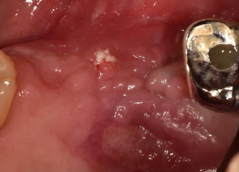
About one week after surgery, some stitches were removed, and at the two-week follow-up, the surgical site showed steady healing.

The Korea Disease Control and Prevention Agency advises avoiding forceful nose-blowing or hard sneezing for about 3-4 weeks after an oroantral communication occurs.
It’s important to follow your care team’s guidance so that excessive pressure isn’t placed on the surgical site.
Why Does Accurate Diagnosis Matter Before and After Closure Surgery?
Having an oroantral fistula doesn’t mean the same treatment is needed in every case.
However, if an opening remains for a long time after an extraction or implant removal, or if a feeling of air passing between the nose and mouth keeps recurring, it’s worth having your current condition checked.
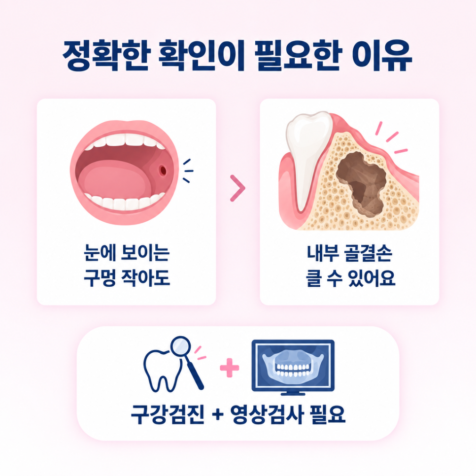
As in this case, even if the opening visible in the mouth is small, the actual extent of internal bone loss can be different.
That’s why, rather than judging by appearance alone, it’s important to check the fistula’s location and extent, the surrounding bone, and the condition of the maxillary sinus together through an oral exam and imaging tests.

My Life Dental Clinic reviews each patient’s oral condition and imaging results comprehensively before discussing the appropriate treatment direction.
If an area doesn’t heal for a long time after an upper molar extraction or implant removal, or if unusual discomfort continues, it’s worth having your current condition checked rather than leaving it untreated.
If you’d like more dental information, you can find additional resources at [My Life Dental Clinic] Home of a Doctor With Clinical Teaching Experience.

Frequently Asked Questions
1. Can an oroantral fistula close on its own?
A small passage in its early stages can sometimes heal naturally, depending on the condition. However, this varies by size, duration, and the state of the maxillary sinus, so if symptoms persist for a long time, it’s necessary to confirm the condition through an exam.
2. If the opening in my mouth looks small, is that okay?
It’s difficult to judge the internal condition based on the visible size of the fistula alone. As in this case, there can be a difference between the opening visible to the eye and the extent of internal bone loss, so imaging tests are performed together when needed.
3. Is oroantral fistula closure surgery always performed the same way?
No, it isn’t. The treatment method can vary depending on the fistula’s location and size, the surrounding tissue and bone loss, and the condition of the maxillary sinus. It’s necessary to assess each individual’s condition and then establish an appropriate treatment plan.
References
· Korea Disease Control and Prevention Agency, “Tooth Extraction,” National Health Information Portal. View source
· Journal of the Korean Dental Association, “Closure of Chronic Oroantral Fistula Through Restoration of the Sinus Membrane,” Korean Dental Association. View source
· ScienceDirect Topics, “Oroantral Fistula – an overview,” ScienceDirect. View source
· Dental Arirang, “[Dental MBA] Oroantral Fistula Closure (Oroantral Fistula closure) Billing Criteria,” Dental Arirang. View source
※ This article is intended for general health information purposes only and is not a substitute for individual diagnosis or treatment. If you have symptoms, please consult a specialist.